
Navigating optimal abdominal CT protocols is crucial; contrast enhancement aids diagnosis significantly, especially when evaluating vessels and organs.
Consider ACR Appropriateness Criteria when ordering.
Effective CT scan ordering requires a thoughtful approach, balancing diagnostic benefits with potential risks. Physicians must judiciously select the appropriate protocol – with or without contrast – based on clinical indication.
Johns Hopkins Medicine emphasizes contrast’s value in visualizing vessels and organs, enabling more accurate diagnoses.
This guide aims to assist referring physicians in making informed decisions, considering factors like renal function and alternative imaging modalities like MRI.
Understanding ACR Appropriateness Criteria is paramount, alongside awareness of radiation exposure and long-term consequences. Prioritizing patient safety and informed consent is essential.
Understanding the Basics of CT Scans
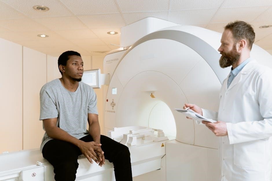
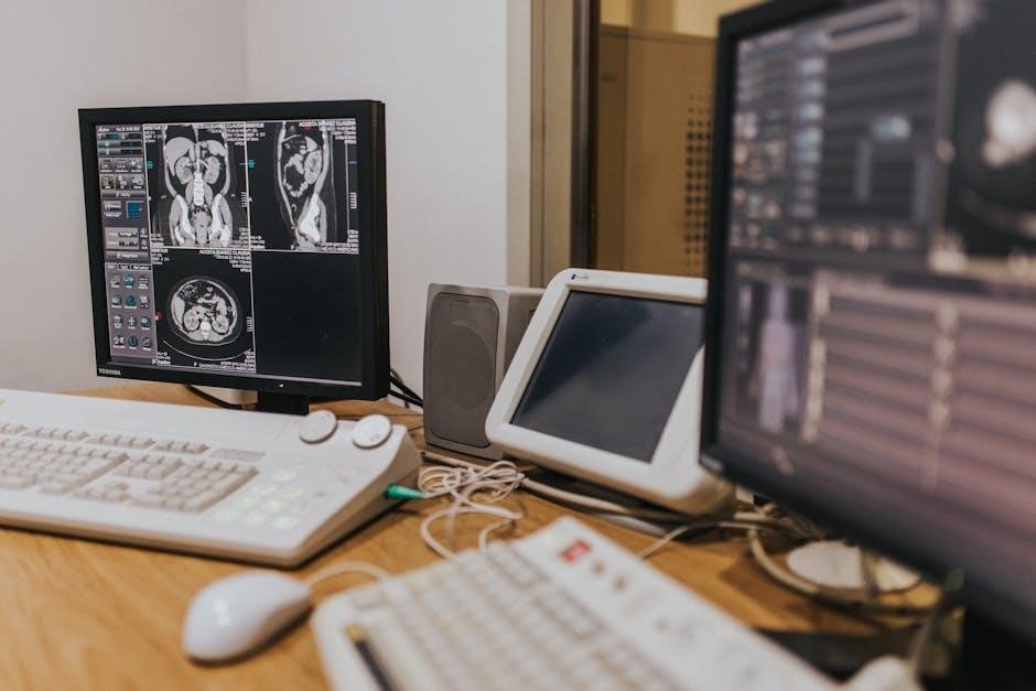
Computed Tomography (CT) scans utilize X-rays to create detailed cross-sectional images of the body. These images reveal structures like organs, bones, and soft tissues with greater clarity than traditional X-rays.
Contrast enhancement, often involving intravenous (IV) contrast, significantly improves visualization of blood vessels and certain organs, aiding in diagnosis.
However, CT scans involve radiation exposure, necessitating careful consideration of the benefits versus risks.
Appropriate scan selection, guided by clinical questions and ACR criteria, minimizes unnecessary radiation. Understanding these basics is fundamental for responsible CT scan ordering.
What is a CT Scan?
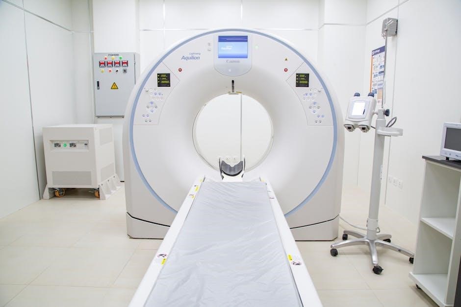
A CT scan, or Computed Tomography scan, is a sophisticated medical imaging technique. It employs X-rays taken from multiple angles around the body, then processed by a computer to generate detailed cross-sectional images.
These images provide a comprehensive view of bones, soft tissues, and blood vessels, far exceeding the detail of standard X-rays.
CT scans are invaluable for diagnosing a wide range of conditions, from internal injuries and infections to cancers and cardiovascular diseases.
The use of contrast agents can further enhance image clarity, revealing subtle abnormalities.
How CT Scans Work
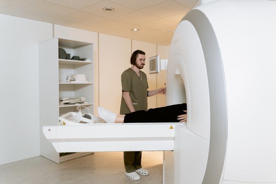
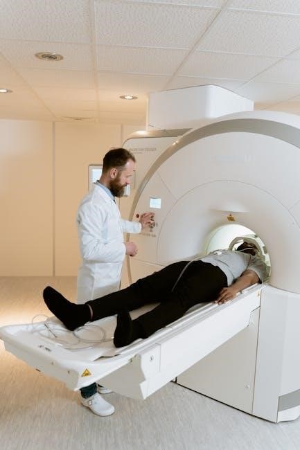
CT scans utilize an X-ray tube that rotates around the patient, emitting a beam of radiation. Detectors positioned opposite the tube measure the amount of radiation that passes through the body. As the tube rotates, numerous images are captured from different angles.
A powerful computer then reconstructs these images into cross-sectional “slices,” creating a detailed visual representation of internal structures.
Contrast agents, when used, enhance the visibility of blood vessels and organs, improving diagnostic accuracy.
Indications for CT Scans
CT scans are invaluable in emergency situations, such as detecting internal bleeding, assessing trauma injuries, and diagnosing stroke. Pre-operative planning for fractures also benefits from CT imaging, providing detailed bony visualization.
Furthermore, CT scans play a vital role in oncology, particularly in colon cancer staging and monitoring response to adjuvant chemotherapy. However, careful consideration of renal function is essential when utilizing contrast-enhanced CT, as impairment necessitates alternative imaging strategies like MRI.
Emergency Situations Requiring CT
Rapid assessment is critical in emergencies, making CT scans a cornerstone of initial evaluation. These scans are essential for identifying internal injuries following trauma, such as fractures or organ damage.
Furthermore, CT excels in detecting acute conditions like stroke, pulmonary embolism, and appendicitis, guiding immediate treatment decisions. Approximately 175 million ED encounters utilize CT head scans annually, highlighting their prevalence. Prompt imaging can significantly impact patient outcomes in these time-sensitive scenarios.
CT for Pre-Operative Planning (Fractures)
CT scans are invaluable for detailed fracture assessment prior to surgical intervention. While initial imaging for bony lesions often begins with X-rays, CT provides superior visualization of complex fractures, particularly those involving joints.
This detailed assessment aids in precise surgical planning, ensuring optimal implant placement and fracture reduction. However, for suspected occult fractures, infection, or osteomyelitis, MRI is generally preferred, offering better soft tissue contrast.
Contrast Enhancement: When is it Necessary?
Contrast enhancement significantly improves diagnostic capabilities in CT scans, particularly for abdominal imaging, allowing radiologists to visualize vessels and organs more effectively. It enables diagnoses often missed on non-contrast scans.
However, contrast isn’t always required. Protocols should be tailored to specific indications. If a patient lacks contraindications to IV contrast, a CT abdomen/pelvis with IV contrast is generally recommended. Careful consideration of renal function is paramount before administering contrast.
Benefits of IV Contrast
Intravenous (IV) contrast dramatically enhances the visualization of blood vessels and internal organs during CT scans. This improved visibility allows radiologists to detect subtle abnormalities that might be undetectable on non-contrast imaging.
Specifically, contrast helps differentiate between normal and abnormal tissues, aiding in the diagnosis of various conditions, including tumors, infections, and vascular diseases. Johns Hopkins Medicine emphasizes its crucial role in making many diagnoses. However, patient safety, particularly renal function, must be carefully assessed.
Abdominal CT Protocols with and without Contrast
Selecting the appropriate abdominal CT protocol – with or without IV contrast – is paramount for accurate diagnosis. Contrast enhancement is invaluable when detailed visualization of vessels and organs is needed, as highlighted by Johns Hopkins Medicine.
However, a non-contrast CT may suffice for specific indications. Generally, if no contraindications to IV contrast exist, a CT abdomen/pelvis with IV contrast is recommended. Careful consideration of patient factors, including renal function, is essential for protocol selection.
Considerations for Patients with Renal Impairment
Patients with impaired renal function require special attention when considering CT scans. The American College of Radiology (ACR) suggests that for a GFR less than 30, a non-contrast MRI is often more informative than a non-contrast CT.

This is due to the risk of contrast-induced nephropathy with IV contrast agents. Thoroughly assess the patient’s GFR before ordering. Alternative imaging modalities, like MRI, should be prioritized to minimize potential harm and ensure optimal diagnostic evaluation.
GFR and CT Scan Ordering
Glomerular Filtration Rate (GFR) is a critical factor when determining the appropriateness of CT scans, particularly those utilizing IV contrast. A GFR below 30 mL/min/1.73m² significantly increases the risk of contrast-induced nephropathy;
Careful consideration must be given to the risk-benefit ratio. When possible, explore alternative imaging options like non-contrast MRI. If CT with contrast is deemed necessary, hydration protocols and minimizing contrast dose are essential to protect renal function.
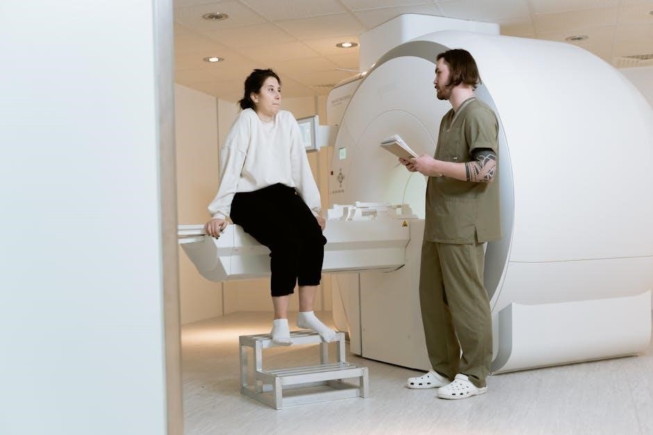
MRI as an Alternative for Impaired Renal Function
For patients exhibiting impaired renal function – specifically a GFR less than 30 – MRI often presents a safer and more informative alternative to non-contrast CT scans. Unlike CT with contrast, MRI doesn’t carry the same risk of inducing nephropathy.
MRI can effectively visualize soft tissues and detect abnormalities without relying on iodinated contrast agents. Utilizing MRI, with and without contrast, is particularly beneficial when evaluating occult fractures, infections, or osteomyelitis.
CT Scans for Musculoskeletal Issues
Initial imaging for suspected bony lesions should consistently begin with X-ray; this provides a foundational assessment. However, when occult fractures are suspected, or when investigating potential infection like osteomyelitis, MRI becomes the preferred modality.
MRI, utilized both with and without contrast, offers superior soft tissue detail, crucial for identifying subtle fractures or assessing the extent of infection. CT scans play a role in pre-operative fracture planning, complementing the information gained from X-ray.
Initial Imaging for Bony Lesions (X-ray)
For the primary evaluation of bony lesions, X-ray remains the cornerstone of initial imaging. It’s a readily available, cost-effective, and efficient method for detecting fractures, dislocations, and certain bone abnormalities.
However, X-ray’s limitations in visualizing soft tissues necessitate further investigation with more advanced modalities like MRI or CT scan when subtle injuries or complex pathology are suspected. Always prioritize X-ray first before considering more radiation-intensive options.
MRI for Occult Fractures, Infection & Osteomyelitis
When X-rays are inconclusive, MRI excels at detecting occult fractures—those not visible on standard radiographs. Its superior soft tissue contrast reveals bone marrow edema, a hallmark of hidden fractures.
Furthermore, MRI is invaluable in diagnosing bone and soft tissue infections, including osteomyelitis. MRI, with and without contrast, differentiates between inflammation and infection, assessing the extent of disease. It’s crucial for guiding treatment decisions and monitoring response in these complex cases.
CT Scans and General Pain
CT scans are generally not the first-line imaging modality for evaluating general, nonspecific pain. While CT can identify structural abnormalities, its use should be carefully considered due to radiation exposure.
For patients presenting with general pain, MRI often provides a more comprehensive assessment without ionizing radiation. MRI excels at visualizing soft tissues, ligaments, and tendons, which are frequent sources of pain. Utilizing MRI without contrast is often the preferred initial approach for these cases, offering detailed anatomical information.
Utilizing MRI for General Pain Assessment
MRI is a superior choice for assessing general pain, offering detailed soft tissue visualization without ionizing radiation. It’s particularly valuable for evaluating ligaments, tendons, and muscles – common pain generators.
Consider MRI without contrast as the initial imaging modality. If initial MRI findings are inconclusive, or specific pathology is suspected (infection, osteomyelitis), MRI with and without contrast may be warranted. This approach minimizes radiation exposure while maximizing diagnostic yield for complex pain presentations.
Radiation Exposure and Long-Term Risks
While CT scans are invaluable diagnostic tools, awareness of radiation exposure is paramount. Mounting evidence suggests potential long-term consequences, necessitating careful consideration before ordering.
Discuss these risks with patients, ensuring informed consent. Minimize exposure by adhering to the “As Low As Reasonably Achievable” (ALARA) principle and utilizing appropriate imaging protocols. Explore alternative modalities like MRI when feasible, particularly for repeat imaging or in younger patients, to mitigate cumulative radiation dose.
CT Head Scan Usage Statistics
CT head scans represent a significant portion of diagnostic imaging utilization. Approximately 175 million emergency department (ED) encounters involved CT head scans, highlighting their frequent application in acute care settings.
This high volume underscores the need for judicious ordering practices. Consider clinical necessity and alternative imaging options to minimize unnecessary exposure. Staying informed about appropriate utilization guidelines and regional benchmarks can help optimize patient care and reduce overall scan rates, improving resource allocation.
CT Scans in Oncology: Colon Cancer
CT scans play a vital role in managing colon cancer, particularly in assessing treatment response and detecting recurrence. The TAB-EOAO study investigated trends in adjuvant chemotherapy utilization based on colon cancer onset age.
Understanding the benefits of chemotherapy alongside appropriate CT imaging is crucial. As presented in a 2025 ASCO meeting abstract, analyzing these factors can refine personalized treatment strategies. CT scans help monitor disease progression and guide clinical decision-making throughout the oncology care pathway.
Adjuvant Chemotherapy and CT Scan Utilization
The interplay between adjuvant chemotherapy and CT scan utilization is a key area of focus in colon cancer management. The TAB-EOAO study specifically examined trends in chemotherapy use, correlating it with early versus average-onset locally advanced colon cancer.
CT scans are essential for monitoring treatment response to chemotherapy, detecting potential recurrence, and assessing overall disease burden. Analyzing these imaging results informs adjustments to the chemotherapy regimen and contributes to improved patient outcomes. Careful consideration of both modalities is paramount.
ACR Appropriateness Criteria
Adhering to the American College of Radiology (ACR) Appropriateness Criteria is vital for responsible CT scan ordering. These guidelines provide evidence-based recommendations, optimizing imaging selection for specific clinical scenarios.
For patients with impaired renal function – specifically a GFR less than 30 – the ACR suggests a non-contrast MRI is often more informative than a non-contrast CT. Utilizing these criteria minimizes unnecessary radiation exposure and ensures appropriate image acquisition, leading to improved diagnostic accuracy and patient safety.
Ordering CT Scans: A Summary for Referring Physicians
When considering abdominal CT imaging, prioritize contrast-enhanced scans unless contraindications exist. Contrast significantly improves diagnostic capability by highlighting vessels and organs, revealing details unseen on non-contrast studies.
For pre-operative fracture planning, CT scans are valuable. However, for occult fractures, infection, or osteomyelitis, MRI is generally preferred. Remember to assess renal function; if GFR is below 30, explore MRI alternatives to minimize risk. Stay informed about updated research and guidelines for optimal patient care.
Staying Updated on New Articles & Research
Continuous learning is vital in the rapidly evolving field of medical imaging. Approximately 175 million ED encounters involved CT head scans, highlighting their frequent use. New research continually refines protocols and assesses long-term risks associated with radiation exposure.
Stay abreast of publications like those from ASCO, such as the TAB-EOAO study on adjuvant chemotherapy in colon cancer. Provide your email to receive updates on new articles. Utilizing resources like the American College of Radiology’s Appropriateness Criteria ensures evidence-based ordering practices.
Expertise in Health and Medicine
A strong foundation in health and medicine is paramount for informed CT scan ordering. Professionals like Laura, holding a Masters in Experimental Neuroscience and a Biology Bachelor’s from Imperial College London, exemplify this expertise.
Understanding the nuances of imaging modalities, including CT and MRI, is crucial for appropriate utilization. This knowledge extends to recognizing indications for contrast enhancement and considering alternative options for patients with renal impairment. Effective patient communication, coupled with medical proficiency, ensures optimal care.
Patient Communication and Informed Consent
Open communication with patients regarding CT scans is essential. Patients need to understand both the benefits – detecting injuries and illness – and the potential long-term consequences, including radiation exposure.
Informed consent should encompass a discussion of risks and alternatives, such as MRI, particularly for those with impaired renal function. Transparency builds trust and empowers patients to participate actively in their healthcare decisions. Clearly explaining the rationale for the scan and potential findings is vital for a positive patient experience.